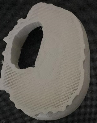
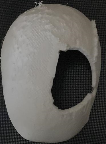
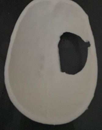
Introduction: In the world of medical imaging, DICOM (Digital Imaging and Communications in Medicine) has emerged as a standard format for storing and transmitting medical images. However, the true potential of DICOM goes beyond mere 2D images. With advancements in technology, the conversion of DICOM data into immersive 3D structures has revolutionized medical visualization, leading to enhanced diagnostics, treatment planning, and surgical precision. In this article, we explore the remarkable journey of transforming DICOM images into captivating 3D structures and the impact it has on the medical field.
Enhanced Visualization: DICOM to 3D structure conversion allows healthcare professionals to transcend the limitations of traditional 2D imaging. By reconstructing volumetric data from DICOM images, intricate anatomical structures can be visualized in three dimensions, providing a comprehensive understanding of complex medical conditions. This improved visualization aids in precise identification, measurement, and assessment of abnormalities, promoting accurate diagnosis and treatment decisions.
Precise Treatment Planning: The ability to convert DICOM images into 3D structures has significantly influenced treatment planning processes. Surgeons can now simulate procedures and visualize the patient’s anatomy from multiple angles, allowing them to develop intricate surgical strategies and anticipate potential challenges. This preoperative insight enhances precision, reduces risks, and optimizes patient outcomes. Furthermore, with 3D models, surgeons can better communicate with patients, explaining treatment options and potential outcomes in a more comprehensive and understandable manner.
Medical Education and Research: DICOM to 3D structure conversion has become an invaluable tool in medical education and research. Medical students can gain a deeper understanding of anatomical structures by exploring realistic 3D representations derived from DICOM images. Additionally, researchers can utilize 3D models to study the progression of diseases, evaluate treatment effectiveness, and develop new therapeutic approaches. These advancements contribute to the overall growth of medical knowledge and the improvement of patient care.
Collaboration and Communication: The conversion of DICOM images into 3D structures enhances collaboration and communication among healthcare professionals. By sharing interactive 3D models, physicians, radiologists, and surgeons can collaborate seamlessly, discuss cases, and exchange valuable insights. This collaborative approach leads to more accurate diagnoses, refined treatment plans, and ultimately, better patient outcomes. Furthermore, the ability to present 3D models to patients fosters informed decision-making, empowering individuals to actively participate in their healthcare journey.
Future Possibilities: As technology continues to evolve, the potential applications of DICOM to 3D structure conversion are expanding. Artificial intelligence (AI) and machine learning algorithms can be integrated into the conversion process, enabling automated segmentation and enhanced accuracy. Furthermore, the integration of virtual reality (VR) and augmented reality (AR) technologies with DICOM data holds the promise of creating immersive experiences, allowing healthcare professionals to interact with 3D structures in real-time.
Conclusion: The transformation of DICOM images into 3D structures has paved the way for a new era in medical visualization. This advancement offers enhanced visualization, precise treatment planning, improved medical education and research, enhanced collaboration, and future possibilities for even more innovative applications. As the integration of technology and medicine continues to evolve, DICOM to 3D structure conversion stands as a remarkable tool that empowers healthcare professionals, improves patient outcomes, and revolutionizes the way we understand and approach medical imaging.


